3-Year Checkup Results…Sort Of

I turned 62-years old on December 22nd. How did I celebrate? Well, we had a fantastic dinner in Portland, then I had an MRA next door in Scarborough. Not how everyone would want to celebrate, but it is what it is. That just happened to be the time and date they had available for the MRA, so I took it.

I wasn’t nervous regarding the MRA itself, other than remaining still during the painless, but noisy procedure, it’s always the impending results where the scanxiety comes in to play.

Usually there is a period of waiting before finding out the results, but in this case, the results were posted to the hospital’s online portal the following morning. I wasn’t expecting to see them so soon. There was good news, and potentially some bad news.

The good news is that no new brain aneurysms were detected. That IS good news because with two previous aneurysms, I am already susceptible to develop more.

The bad news may NOT be bad news, but I don’t know that yet. The wording in the report said there is “a 4 mm focus of flow related enhancement” somewhere along the coiled aneurysm. When the doctor’s office called the next day, they said Dr. Ecker said he couldn’t tell if that finding was “real or not” and it appeared he was leaning towards my having a cerebral angiogram to confirm or deny that finding.

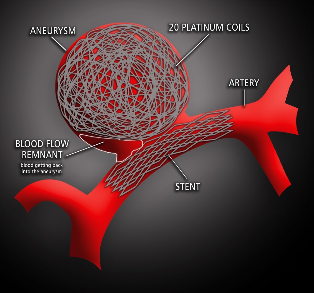
What IS discerning about the 4 mm area of focus is that after my last scan in 2022, the older remnant area of concern where blood was getting back into the neck of my coiled aneurysm, was almost completely gone. So, this is a NEW area of concern. And if it is truly a new out-pouching of blood along the aneurysm, it certainly developed quickly.

Images from before flow-diverter stent and after (Dec. 2022)

We’re hoping to speak to the doctor via TeleHealth, so we don’t have to drive 60 miles just to spend 15 minutes with him, but we DO want to see the images and discuss it with him. I’m hoping that conversation happens sooner rather than later and an angiogram can be scheduled fairly quickly. I want to know if this is “real or not”. If it’s just a blip on the MRA scan, then great, nothing to worry about. If it IS a new remnant, the next course of action could be very complicated as there are already 20 coils and two stents trying to stop this bugger from killing me.

I will update you all when I know more. I’m trying not to be overly reactive, but it certainly is concerning news…until I learn it’s NOT concerning news. Happy birthday to me!


I shared my story (up to this point) on the MaineBA.org website if you’re new to my blog and would like the condensed version of my story.

The Thing that Won’t Go Away

The follow-up with my interventional radiologist/neurosurgeon confirmed the MRA I had in December showed additional development of more blood getting into the neck of my first brain aneurysm – AGAIN. We went through these discussions back in 2015 when that remnant first showed up. I decided to kick the can and watch, wait, and continue to monitor it. After five years, it appears there is more blood now.

The doctor wants me to have another angiogram to get a closer look and determine if we need to take action now. This is not the way I had hoped to start the new year. It may be nothing alarming…or it might be.

I created the graphic below to present, in it’s simplest form, what’s happening. This is the first aneurysm that ruptured in 2006. It’s 11mm or about 1/2” in diameter.

  • There were 16 coils inserted into the aneurysm in 2006.
  • Those coils healed and clotted, preventing blood from getting back into the aneurysm.
  • Five years later, the coils either started to compact, the shape of the aneurysm and neck of the aneurysm changed, or a combination of both occurred, This allowed some blood to get back into the neck of the aneurysm.
  • 4 more coils were inserted (for a total of 20) and a stent was placed across the bottom of the neck in the artery in 2011.
  • In 2015, during annual checkups, it was discovered more blood was starting to get back into the aneurysm again.
  • We decided to “wait and watch” the remnant of blood for future development.
  • Now in 2022, we’re discussing the fact that it appears MORE blood is getting into the aneurysm after six years of stability. This is also a brain aneurysm that has already ruptured once.

During the last 15 years, I have had to endure a couple of “watch and wait” periods for both my aneurysms. There is always a nagging worry that lays there asking those probing questions: Is it growing? Has there been any change? Am I making it worse? Could something I’m doing make it rupture? Is that headache a sign that something is wrong?

Anyone who is living with brain aneurysms knows that worry. Telling me to “Not worry about it” is far easier said than done.

The good news is that I feel fine. I just hope I can stay that way. 🤞

MRA Results

I have added some new images to my brain photography exhibition after my latest MRA last week to check my vascular health. I haven’t had one in two years, so the scan-xiety built up a little bit the day & evening before. Not that I was nervous about the MRA, I have had plenty of those (see the previous blog post!) but it’s always the results that create that uncertainty.

I was especially worried about this one because I haven’t been taking care of myself the last two years. I’m not exercising, I’m not eating right, and since my last MRA in 2018 I had developed high blood pressure. All three of those things are NOT good for maintaining good blood flow in the brain…or elsewhere for that matter. I take full responsibility…no excuses other than being lazy.

I have been reticent to sign up for all the online medical charts offered by the hospitals lately, but I decided to sign up for the one used by the state. As a result, I could view the initial findings of the MRA online on a Sunday before the doctor’s office ever called me on a Tuesday. I’m still debating whether that is a good thing to have or a bad thing. If the results have come back badly, I’m not sure how I would have handled that.

THANKFULLY, the findings were positive and my doctor’s office confirmed the diagnostic radiologist’s report with his reply and recommendations for follow-up.

Here are the main things that came from the report:

  • There is normal flow within both vertebral arteries. No other aneurysm is identified. There is normal flow within both P1 segments and in the right middle cerebral artery.
  • Stable appearance of the coil embolization and previously clipped left internal carotid artery bifurcation aneurysm. Small amount of flow is unchanged or slightly less apparent in the region of the aneurysm neck that measures between 3 and 4mm in size.

The first point is the best one, indicating no other aneurysm was found. I am at higher risk with my family history and my own history with two already.

The second point was also good news about the bloody remnant (narrow area where blood is starting to creep back into the aneurysm) at the neck of the original brain aneurysm being “unchanged” and that it may be “slightly less apparent”. We have been watching this remnant for several years now and I’m always nervous it will increase in size, or create a weak area and possibly burst.

When the doctor recommended I come back in a year for another MRA, I was surprised it’s not another two year follow-up. So, I have sent in a question asking why.

Celebration Tea

I am blessed to still be here and share my story with others. Keep on living. I am enjoying a celebratory scone with tea to celebrate!

I Am Lit

As I get prepared for my 2-year follow-up MRA this Friday, I was curious to see just how many angiograms and MRA/MRI’s my brain has had since 2006. Thankfully, I was able to get a pretty good handle on that number by simply searching this blog. I was surprised by the number…but not really.

CEREBRAL ANGIOGRAMS

I have had NINE cerebral angiograms which include three treatments that are performed using this same process: my initial coiling, then my stent placement, and recoiling were all performed using angiography.

A Cerebral angiography provides x-ray images of blood vessels in and around the brain, showing abnormalities like brain aneurysms.

PHOTO: John Hopkins Medicine

Typically a catheter is inserted into the femoral artery in the groin and threaded through the circulatory system to the carotid artery where contrast die is injected. A series of images are taken as the contrast agent spreads through the brain’s arterial system, then a second series as it reaches the venous system.

MRA/MRI

I have also had SEVEN MRA/MRI’s. Magnetic resonance angiography–also called a magnetic resonance angiogram or MRA–is a type of MRI that looks specifically at the body’s blood vessels. Unlike an angiogram, which requires inserting a catheter into the body, magnetic resonance angiography is a far less invasive and less painful test.


PHOTO: My brain bling via MRA imaging.

The frequency and type of follow-up procedures I receive hinge on the previous scan showed and what procedure was provided. We’re hoping I can continue just getting MRA’s, but if something unusual shows up, more angiograms may be needed. Angiograms are more invasive and risky, but they are the gold standard for getting the clearest images of my blood vessels.

I am blessed to have had such a great neuro team here in Maine and terrific health insurance coverage through work.

Here’s to more scanxiety.