
So…it turns out my new infundibulum is actually an old infundibulum and has been around since at least 2022, possibly before then. It IS visible in my 2022 angiogram images and it’s mentioned in the report, which I didn’t check out until we got home. It wasn’t really discussed then that I can remember. At that time, we were mainly concerned about the flow diverter and if it had stopped the blood from gathering at the neck of my original brain aneurysm — which it has.
Dr. Ecker explains this particular infundibulum is not threatening, it’s stable, and we will continue to watch it over the years. It is filled with blood, but we’re not sure why it showed up as a NEW artifact on the MRA. That’s the whole reason I then had the angiogram: to pinpoint what that artifact was.
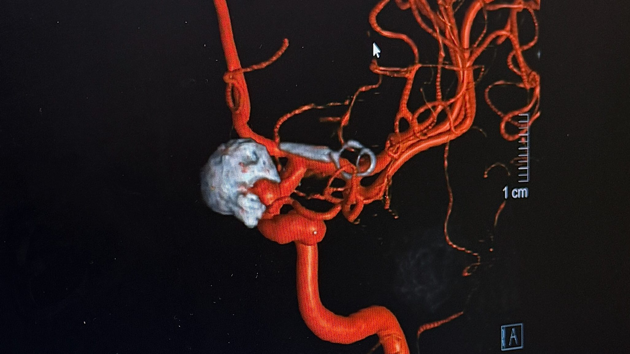
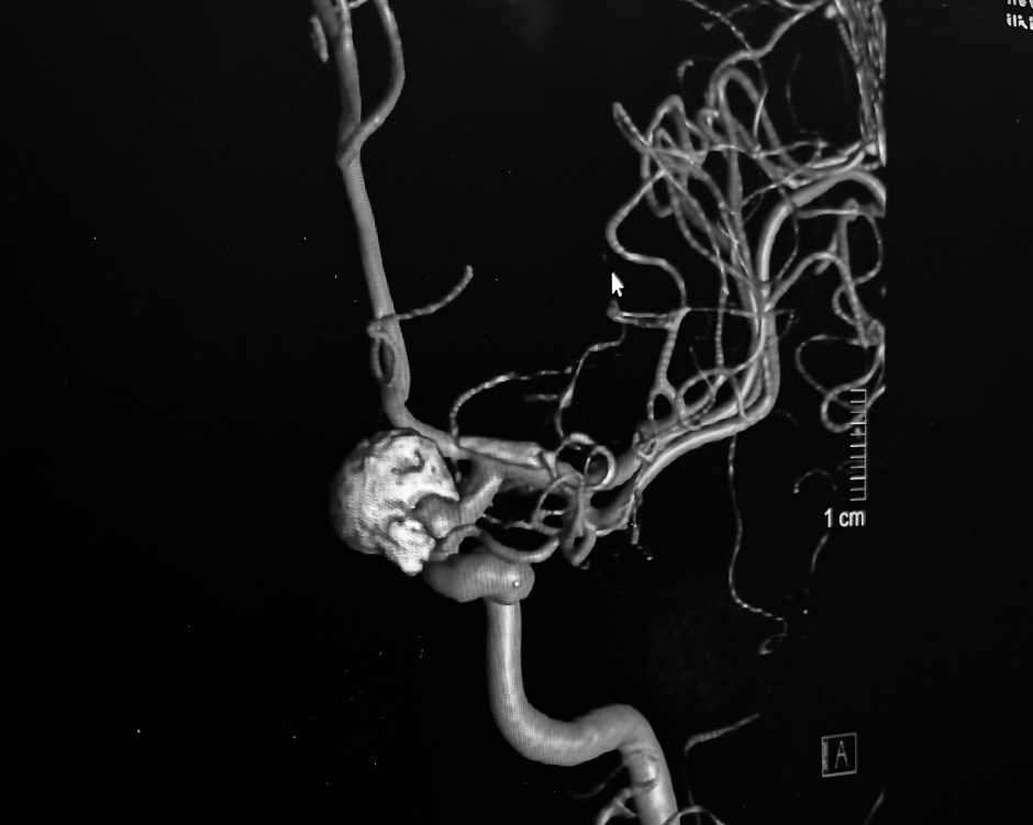
I was able to grab a screen capture of the latest angiogram images in the doctor’s office. Really fascinating to see the large 11mm original aneurysm filled with coils showing as a large gray mass. Then to see how close the clip actually is to the first aneurysm. As Dr. Ecker said, neurologically speaking “it’s in another room.” due to the microscopic aspect of their work. He also pointed out where the infundibulum is. It looks big to me, but it’s only around 3.7mm.
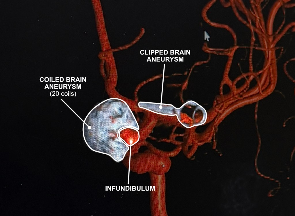
So, the good news is that’s it’s nothing new, but it’s there. Am I happy something is there? No. Not really. Just one more thing to monitor. The plan now is to do a follow-up MRA in two years.
Now that I have learned how to pronounce infundibulum, I hope that’s all I have to be concerned with.
Discover more from Heidi's Brain Blog
Subscribe to get the latest posts sent to your email.

I am so relieved to read that this is actually an old infundibulum and it is stable. I can imagine the relief that both you and Uncle Dave felt when receiving these results. I really appreciate you sharing these photos so that I can understand just where it is at. What wonderful news!!!! I was sending up prayers for these results for you. Love you ❤
LikeLike